
Table of Contents
Cardiovascular diseases (CVDs) remain one of the leading causes of death in Australia, making early and accurate diagnosis crucial for effective treatment. An echocardiogram is one of the most valuable diagnostic tools used by healthcare providers to assess heart function and detect cardiovascular conditions.
This non-invasive, ultrasound-based imaging test provides detailed information about the heart’s structure, function, and blood flow, helping doctors diagnose a wide range of heart problems.
In this article, we will explore how echocardiograms contribute to diagnosing various cardiovascular conditions, the procedures involved, the types of echocardiograms, and the role they play in heart disease management.
What is an Echocardiogram?
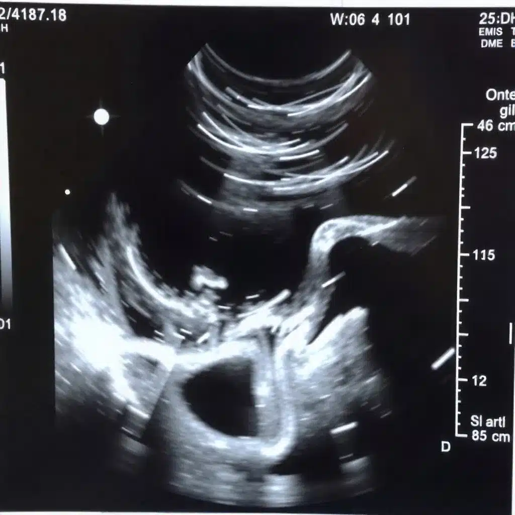
An echocardiogram is a diagnostic procedure that uses high-frequency sound waves (ultrasound) to produce real-time images of the heart. The sound waves are emitted through a transducer, a small device placed on the chest, and then bounced back after hitting the heart’s structures.
These sound waves create detailed images that show the size, shape, and movement of the heart, as well as how the heart chambers and valves function.
Echocardiograms are essential because they allow healthcare providers to observe the heart in action without the need for surgery or invasive procedures. The images provide critical insights into a patient’s heart health and can identify potential problems that may not be detected through other diagnostic methods, such as X-rays or CT scans.
How Does an Echocardiogram Work?
The echocardiogram procedure is quick, non-invasive, and generally painless. Here is how it typically works:
Preparation: The patient is usually asked to lie on an examination table, and a gel is applied to the chest to help the transducer make good contact with the skin.
Image Capture: The transducer is moved over the chest area, emitting sound waves and capturing the reflections. These sound waves create images of the heart’s structures, such as the chambers, valves, and blood vessels.
Assessment: The healthcare provider reviews the images in real-time to assess the heart’s condition and detect any irregularities.
Results: Once the procedure is complete, the results are reviewed, and a diagnosis is made based on the findings.
Depending on the patient’s condition, the healthcare provider may also recommend additional tests, such as a stress echocardiogram, to assess how the heart performs during physical exertion.
Types of Echocardiograms
There are several types of echocardiograms, each providing different perspectives and details about the heart’s function. The type of echocardiogram performed will depend on the symptoms and the information needed by the healthcare provider.
Transthoracic Echocardiogram (TTE)
The transthoracic echocardiogram (TTE) is the most common type and is performed by placing a transducer on the chest. This is typically the first choice for assessing heart function and detecting abnormalities, as it provides clear images of the heart chambers, valves, and blood vessels.
Transesophageal Echocardiogram (TEE)
In cases where the transthoracic echocardiogram does not provide enough information, a transesophageal echocardiogram (TEE) may be used. This test involves placing a specialised probe down the esophagus to capture images of the heart from a closer and clearer angle.
It is particularly useful for detecting problems with the heart valves, blood clots, and other conditions that might be missed with a TTE.
Stress Echocardiogram
A stress echocardiogram is a specialised test used to evaluate how the heart performs under stress, typically induced by exercise or medication. It is helpful for diagnosing coronary artery disease (CAD), which occurs when the heart’s blood supply is reduced due to blockages in the coronary arteries.
Doppler Echocardiogram
A Doppler echocardiogram is used to measure the flow of blood through the heart’s chambers and blood vessels. This technique provides insight into blood velocity, direction, and volume, helping detect abnormal blood flow, which may indicate conditions like valve disease or heart failure.
How Echocardiograms Help Diagnose Cardiovascular Conditions
Echocardiograms play a critical role in diagnosing a variety of cardiovascular conditions. By providing a detailed view of the heart’s structure and function, these tests help healthcare providers detect abnormalities that might indicate serious heart issues.
1. Heart Valve Disease
One of the most common conditions diagnosed with an echocardiogram is heart valve disease. The heart contains four valves—mitral, aortic, tricuspid, and pulmonic—that control the flow of blood through the heart.
If any of these valves become damaged or diseased, they can lead to heart problems such as regurgitation (when blood leaks backward) or stenosis (when the valve doesn’t open fully). An echocardiogram can help detect valve defects, enabling early intervention and proper treatment.
2. Congenital Heart Defects
Many heart defects are present from birth (congenital). An echocardiogram is essential for diagnosing congenital heart conditions such as septal defects (holes in the heart’s walls), malformed valves, or abnormal heart chambers.
Early diagnosis allows for timely treatment, which can significantly improve the patient’s quality of life.
3. Heart Failure
Heart failure occurs when the heart is unable to pump blood effectively, leading to fluid buildup and decreased oxygen supply to vital organs. An echocardiogram helps determine the heart’s pumping efficiency by measuring the ejection fraction (the percentage of blood pumped out of the heart with each beat).
Low ejection fraction readings often indicate heart failure, guiding treatment strategies.
4. Coronary Artery Disease (CAD)
CAD, which involves the narrowing or blockage of the coronary arteries, is one of the most common causes of heart attacks. While a stress echocardiogram is often used to assess CAD, standard echocardiograms can also help identify abnormal heart function or damage caused by reduced blood flow.
This allows healthcare providers to monitor the progression of CAD and recommend appropriate treatments such as angioplasty or bypass surgery.
5. Aneurysms
An echocardiogram can help detect the presence of aneurysms, particularly in the aorta (the large artery that carries blood from the heart to the rest of the body). An aneurysm occurs when the wall of a blood vessel weakens and bulges, potentially leading to rupture.
Detecting an aneurysm early allows for timely intervention to prevent life-threatening complications.
6. Pericardial Disease
The pericardium is the sac surrounding the heart. Inflammation or fluid buildup in this area can cause pericardial disease, which may lead to heart discomfort, chest pain, or breathing difficulties.
An echocardiogram helps visualise the pericardium, allowing healthcare providers to identify any abnormalities and recommend appropriate treatment.
7. Cardiomyopathy
Cardiomyopathy refers to diseases of the heart muscle that affect its ability to pump blood efficiently. There are different types of cardiomyopathy, including dilated, hypertrophic, and restrictive cardiomyopathy.
Echocardiograms help assess the heart’s size, thickness, and function, allowing for early diagnosis and treatment options.
The Benefits of Echocardiograms in Heart Disease Management
Echocardiograms offer numerous benefits when it comes to diagnosing and managing cardiovascular conditions. Some key benefits include:
1. Non-Invasive and Safe
Echocardiograms are non-invasive procedures that do not require surgery or the use of contrast agents, making them a safe and comfortable option for patients. They are also free of radiation, making them ideal for repeated use over time.
2. Real-Time Results
An echocardiogram provides real-time images, allowing healthcare providers to immediately assess the heart’s condition and make quick decisions about treatment. This is particularly beneficial in emergencies or for monitoring the effectiveness of ongoing treatment.
3. Early Detection
Echocardiograms allow for the early detection of heart conditions, often before symptoms arise. This early diagnosis is essential for implementing preventive measures or starting treatment before the condition worsens.
4. Accurate Assessment
Echocardiograms provide detailed images that help healthcare providers accurately assess the heart’s function and structure. This ensures a comprehensive understanding of the patient’s cardiovascular health, leading to better-targeted treatments.
Conclusion
Echocardiograms are an essential tool in diagnosing and managing cardiovascular conditions in Australia. They offer a safe, non-invasive way to obtain detailed, real-time information about the heart’s structure and function, allowing healthcare providers to detect issues early and make informed decisions about treatment.
Whether assessing heart valve disease, congenital disabilities, heart failure, or coronary artery disease, an echocardiogram can significantly improve the accuracy of diagnosis and patient outcomes. With its ability to detect a wide range of heart issues, this diagnostic test plays a critical role in improving heart health and preventing serious cardiovascular complications.